Brain protein changes in mice differ from human Alzheimer’s
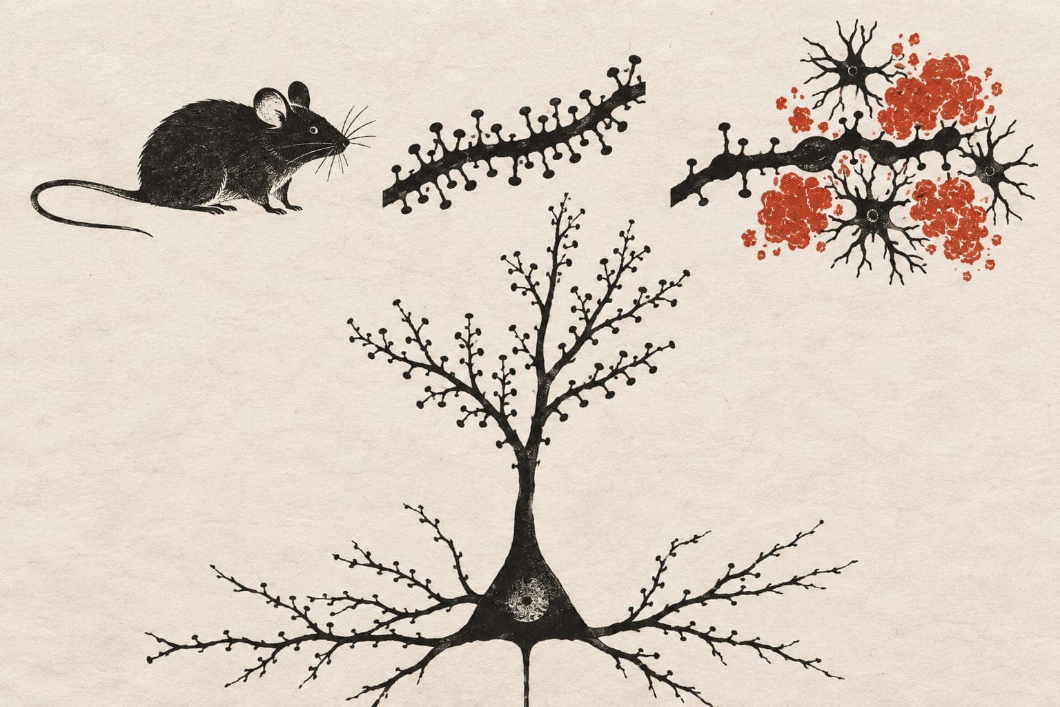
An experiment in six Alzheimer’s model mice and six control mice at 3 and 6 months suggested protein changes differed from human disease.
Our read
Mechanisms- Studied:
- Experiments in 6 mice
- In the record:
- One of 2,836 papers on APP on record: 12 with results in people, 107 in animals.
- Why it’s aging research:
- It studies organisms or tissues that aged naturally.
Editor of record: Boris Djordjevic · AgingResearch.News
AppNLGF mice, a model of Alzheimer’s disease, and genetically unmodified control mice were compared in an animal experiment. Researchers measured protein amounts in two brain regions—the cortex and hippocampus—from six mice of each type at 3 and 6 months.
Differences between model and control mice were extensive. At 6 months, the cortex had 763 protein changes, compared with 304 in the hippocampus. Many changes were already present at 3 months. Comparing ages within each mouse type revealed relatively few changes. The researchers compared disease-associated biological processes with an earlier analysis of the same regions in human Alzheimer’s brains collected after death. The differences suggested that the mouse response to amyloid protein deposits differed substantially from that in human Alzheimer’s disease.
Why it matters
Mouse models help researchers examine early Alzheimer’s-related changes that are difficult to study in human brain tissue. The authors urged careful characterization and selection of these models.
Caveats
The human comparison relied on previously analyzed brain tissue collected after death. The mouse results covered one model, two brain regions and ages of 3 and 6 months.
The paper
Cardiff University; the University of Manchester
Brain Communications, Nov 6, 2026, CC BY

Comments
Be the first to comment.