Brain cells responded less flexibly in Alzheimer’s model mice
An imaging experiment in behaving mice found higher baseline brain-cell activity but weaker movement-driven responses in a brain region involved in memory.
Our read
Mechanisms- Studied:
- Experiments in behaving 5xFAD mice
- What changed:
- Brain-cell activity varied over a narrower range in Alzheimer’s model mice
- The catch:
- Calcium imaging served as a proxy for neuronal firing
Editor of record: Boris Djordjevic · AgingResearch.News
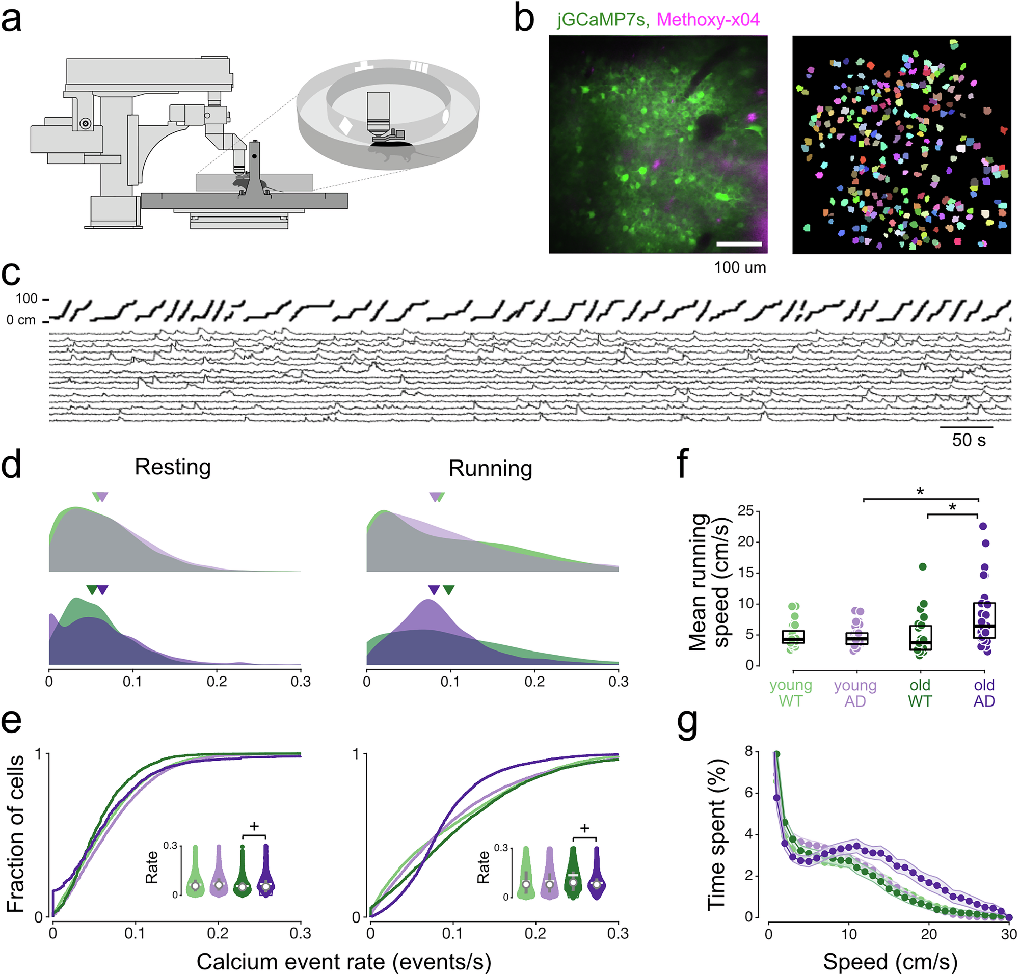
In 5xFAD mice, an Alzheimer’s disease model, an imaging experiment tracked nerve-cell activity in CA1, a memory-related brain area. Cells had higher baseline activity but less activity driven by movement. This narrowed the range over which their activity changed. These abnormalities were strongest near amyloid plaques, clumps of protein, and became more widespread with age.
The researchers also found changes in how cells synchronized their activity, less accurate coding of location and more variable responses. Location-specific activity patterns emerged more slowly in both familiar and new environments, which the authors interpreted as impaired recall and learning. The authors linked local plaque pathology to reduced flexibility in nerve-cell activity and progressive problems with spatial memory coding.
Why it matters
Amyloid buildup is a feature of Alzheimer’s disease. The work addresses how it may disrupt the brain circuits that support spatial memory.
Caveats
The experiment used 5xFAD mice; whether the same narrowing of brain-cell activity occurs in people with Alzheimer’s disease remains open.

Comments
Be the first to comment.