Deleting two genes worsened stroke injury in male mice
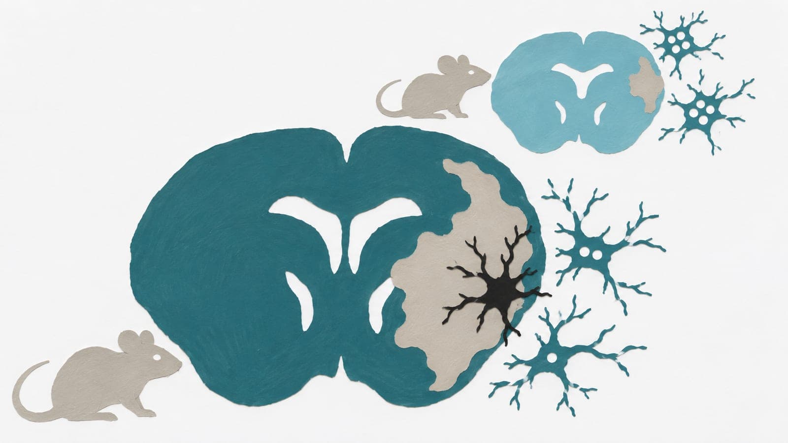
At 48 hours, males lacking the genes in brain immune cells had 2.2-fold larger areas of stroke damage than control males.
Our read
Mechanisms- Studied:
- Preprint: experiments in older male and female mice, 48 hours, against control males
- What changed:
- At 48 hours, males with the gene deletions had 2.2-fold larger areas of stroke damage than control males
- The catch:
- Genes were deleted before stroke; effects of blocking signaling afterward were not measured
Editor of record: Boris Djordjevic · AgingResearch.News
Fifteen-month-old mice underwent an animal experiment modeling stroke caused by blocked blood flow. The researchers deleted two genes involved in calcium signaling in microglia, the brain’s immune cells. They blocked a brain artery and tracked injury with MRI.
Six hours later, mice with and without the deletions had similar areas of stroke injury. From six to 48 hours, these areas expanded 2.7-fold in males with the deletions but did not grow in control males. At 48 hours, females with the deletions did not differ from controls. Imaging also showed that microglial calcium signals were one-third smaller during waves of electrical disruption in the brain.
Why it matters
The findings challenge the idea that dampening microglial calcium signaling limits stroke damage in older mice. The authors suggest this signaling may protect threatened tissue early after stroke, possibly by sustaining blood flow.
Caveats
Deleting genes before an experimentally induced stroke does not show what blocking this signaling after a stroke would do. The preprint has not been peer-reviewed.
The paper
University of Virginia
bioRxiv, Nov 13, 2026, Open access, CC BY, Preprint, not yet peer-reviewed
Comments
Be the first to comment.