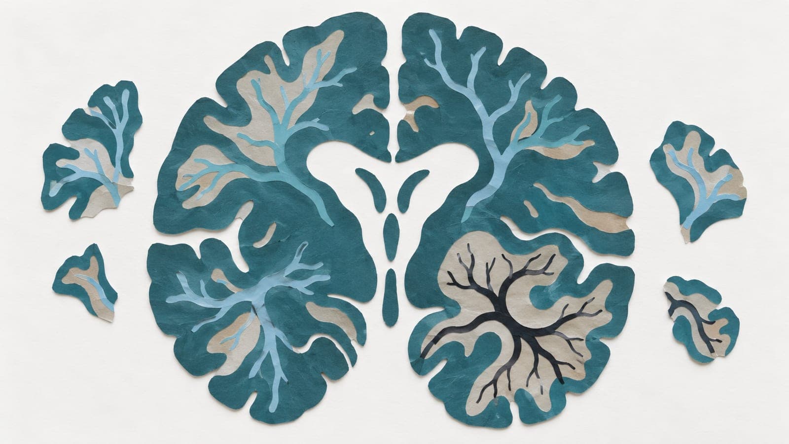
Blood flow and pulses have distinct patterns in human hippocampus
An observational study of 35 young and older adults found age-related differences in blood flow and pulse-related changes in tiny blood vessels.

Limit: The spatial patterns were mapped in only 35 adults
Edited by Boris Djordjevic · AgingResearch.News
In 35 young and older adults, researchers used magnetic resonance imaging in an observational study to map blood flow and pulse-related changes in tiny blood-vessel volume in the hippocampus, a brain region involved in memory. They compared these measurements with reference maps of tissue structure, electrical activity and gene activity.
Blood flow and pulsatility had distinct spatial patterns and age-related differences. In young adults, blood flow corresponded preferentially to the density of one type of nerve cell that dampens other cells’ activity. Blood flow and pulsatility were also associated with different brain-wave patterns. Several associations detected in young adults were not detected in older adults’ maps.
Why it matters
The work offers a framework for investigating how brain circulation relates to nerve-cell activity across aging.
Caveats
The observational design and comparisons with reference maps cannot establish that vascular pulses affect nerve-cell activity. The study included 35 adults and is a preprint, not yet peer-reviewed.
- Mechanotransduction
- Beta/gamma distribution
- Cerebral perfusion
- Microvascular volumetric pulsatility index
- Parvalbumin-interneuron density
- Humans
The paper
University of Southern California
bioRxiv, Oct 21, 2026, Open access, CC BY-NC, Preprint, not peer-reviewed