Osteoporosis therapies affect bone cell spaces differently in mice
In a four-week mouse experiment, a bone-building hormone enlarged the spaces housing bone cells, while a drug that slows bone breakdown stabilised their local structure.
bioRxiv
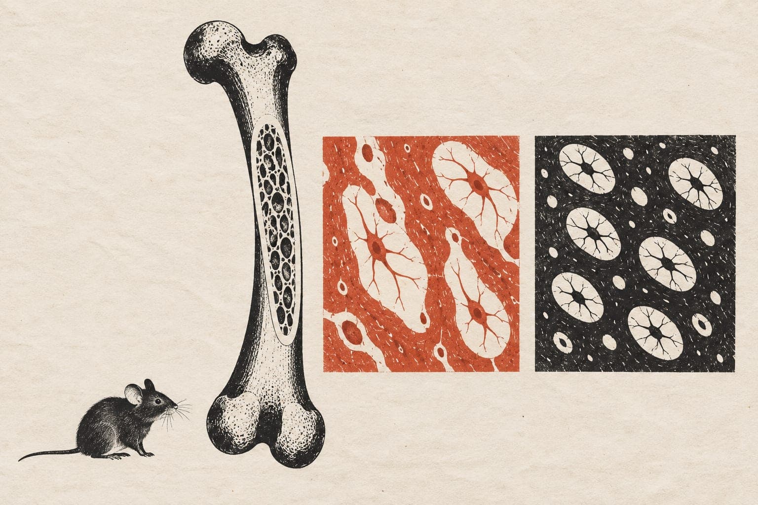
In an experiment in 15-week-old mice with their ovaries removed to model osteoporosis, researchers tested two drugs, alone or with repeated force applied to bone. They gave parathyroid hormone, which promotes bone formation, or a bisphosphonate, which slows bone breakdown, for four weeks. Three-dimensional scans mapped lacunae, the spaces housing bone cells, onto sites where bone formed or broke down.
The bisphosphonate stabilised the local structure of these spaces. Parathyroid hormone enlarged them and made them less spherical, overriding added loading effects. Oestrogen loss made spaces in newly formed bone rounder, disrupting normal differences between bone sites. The therapies restored those differences. However, despite restoring bone volume, they did not fully restore the density of these spaces.
Why it matters
Postmenopausal osteoporosis weakens bone and raises fracture risk. Mapping bone cells’ surroundings may help researchers understand why therapies produce different changes in bone tissue.
Caveats
This was a mouse experiment measuring bone cell spaces, not a study of osteoporosis therapies in people. The report is a preprint and has not been peer reviewed.
- Parathyroid hormone
- Bisphosphonates
- Extracellular matrix remodelling
- Extracellular matrix
- Osteoporosis
- Mice
The paper
Show 4 more authors
F. M. Lambers, F. A. Schulte, G. A. Kuhn, N. Mathavan,ETH Zurich
bioRxiv · 6 Oct 2026 · Preprint, not peer-reviewed