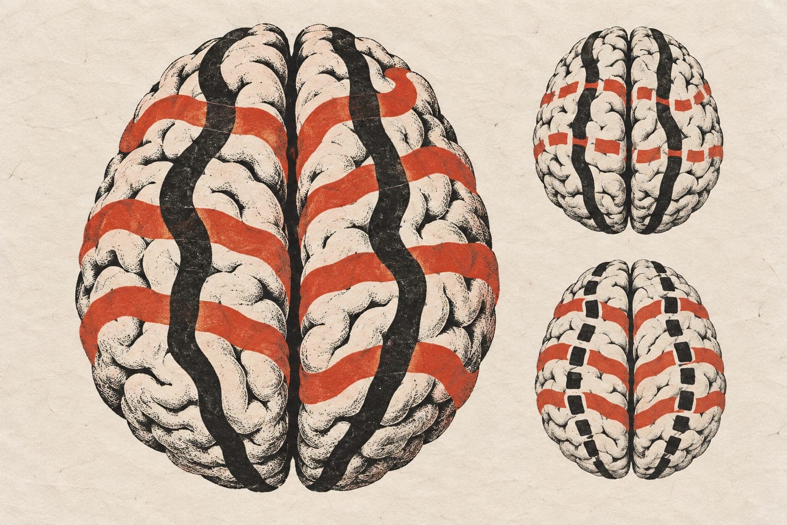
Brain networks changed their relative balance as adults aged
One brain network shifted upward relative to nine others by 5–8% per five years, during rest and three tasks.

Limit: Head motion and scan quality could not be included as covariates
An observational study followed 852 adults aged 36–89 at baseline using repeated functional magnetic resonance imaging, which measures changes in blood oxygenation. Researchers analyzed 2,220 sessions over a median follow-up of 5.5 years. Scans covered rest and tasks involving controlling responses, linking memories, and coordinating vision and movement.
The researchers compared ten networks in the brain’s outer layer. Their relative balance changed over follow-up, differently across states. The frontoparietal network, spanning areas toward the front and upper back of the brain, shifted upward relative to the other nine in all four states. This direction persisted under two ways of adjusting for signal size, although estimates of overall change depended on the adjustment. Researchers detected no within-person link between this relative shift and fluid cognition, the ability to reason through unfamiliar problems.
Why it matters
Following the same people across rest and tasks helps address how brain organization changes within individuals as they age, rather than relying only on differences between younger and older adults.
Caveats
This observational study could not establish cause and effect, and its population did not include people younger than 36 at baseline. The paper is a preprint and has not been peer-reviewed.
From the paper
- Participants
The primary cohort included 852 adults with a mean baseline age of 60.96 years (SD 15.12; range 36–89), of whom 492 (57.7%) were women (Table 1).
Results, 3.1 Overall covariance and signal amplitude declined over follow-up
- Follow-up
Median follow-up was 5.50 years (IQR 3.10–6.33), and 431 participants had at least three sessions.
Results, 3.1 Overall covariance and signal amplitude declined over follow-up
- Limitation
The analysis used released imaging-derived measures, so residual motion and session-level data quality could not be assessed directly, and total amplitude was reconstructed from released products.
Quoted word for word from the full text on arXiv.
- Fluid cognition
- Estimated noise pair amplitude
- Frontoparietal network relative shift
- Functional connectivity
- Native common covariance
- Humans
The paper
arXiv, Oct 17, 2026, Open access, CC BY-NC-ND, Preprint, not peer-reviewed