Dark areas on brain scans showed disease-specific patterns
An observational study compared brain scans from 7,587 patients across 21 brain disorders against an age-based reference built from 34,148 healthy people.

Our read
Biomarkers- Studied:
- Preprint: cross-sectional study of 7,587 patients with neurological or psychiatric disorders (Multicenter)
- What changed:
- Dark areas in white matter showed disease-specific patterns compared with the healthy reference
- The catch:
- Brain disorders could cause the dark areas rather than the other way around
Editor of record: Boris Djordjevic · AgingResearch.News
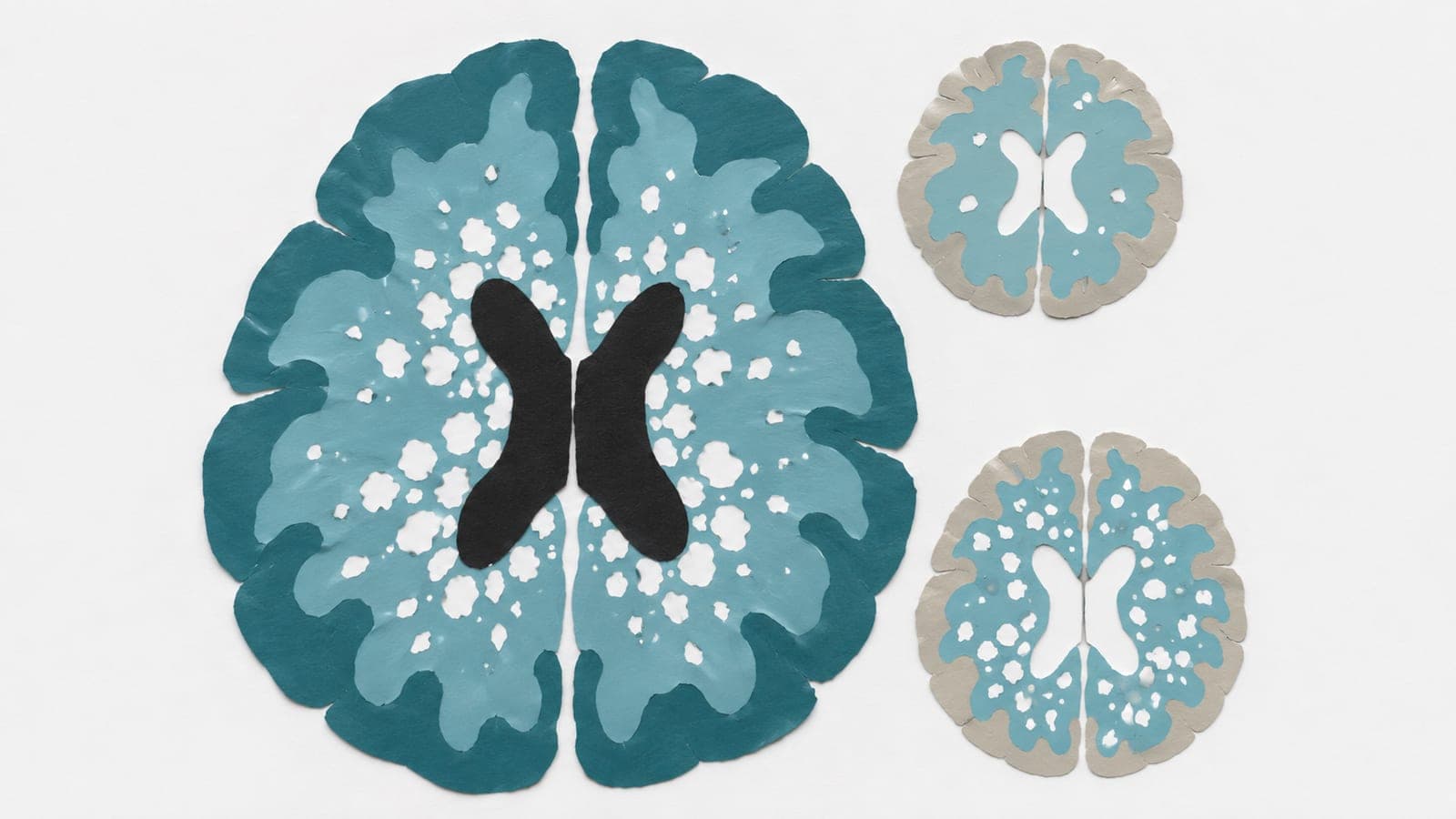
Brain scans from 34,148 healthy people aged 4–85 and 7,587 patients across 21 neurological and psychiatric disorders formed the basis of a multicenter observational study. Researchers measured the volume of dark areas in white matter, the brain’s nerve-fiber tissue, on one type of magnetic resonance imaging scan. They used the healthy group to build an age-based reference.
Patients’ departures from this reference showed disease-specific patterns, with marked differences in neurological diseases. Larger departures were linked to brain shrinkage, cognitive impairment and poorer outcomes. Greater volumes predicted poorer medication response in Parkinson’s disease and faster disability progression in multiple sclerosis. These links remained after accounting for lesions detected by another type of scan.
Why it matters
An age-based reference may help distinguish variation across development and aging from white matter changes linked to brain disorders.
Caveats
The comparisons leave open whether the dark areas contributed to brain disorders or reflected their effects. The report is a preprint and has not been peer-reviewed.
The paper
Capital Medical University
medRxiv, Nov 13, 2026, Open access, CC0, Preprint, not yet peer-reviewed


Comments
Be the first to comment.