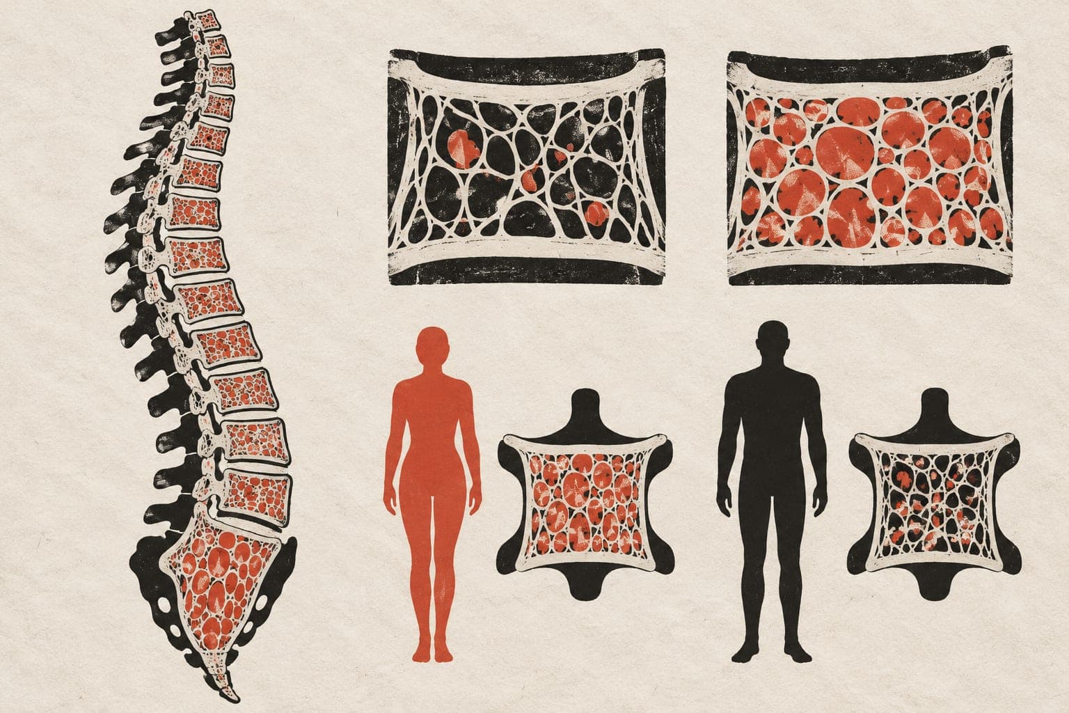
Bone scan readings show different age patterns by sex
Across bone sites, the estimated decrease from age 40 to 80 was a median 34.1% in women versus 12.8% in men.

Multicenter
Limit: Changes in bone readings within individuals were not measured
Edited by Boris Djordjevic · AgingResearch.News
In 78,620 patients aged 15–100 at nine Japanese academic hospitals, researchers examined age and sex patterns in bone scan readings. The retrospective, multicenter cross-sectional study analyzed non-contrast computed tomography (CT) scans taken in 2024, using one scan per patient. Automated software measured attenuation—how much bone weakened the X-ray signal—in 25 spinal bones and 24 ribs.
Readings were lower at older ages at all 49 sites in women and 46 in men. Age patterns differed by sex at every site. The researchers created reference charts comparing each measurement with those of people of the same age and sex. They concluded that measurements from spinal bones adjacent to the first lower-back vertebra may substitute for its measurements, with a stated margin of error; rib measurements may not.
Why it matters
Age-related bone loss raises the question of how to distinguish an unusually low scan reading from one typical for a person’s age and sex.
Caveats
The cross-sectional design compared people of different ages rather than tracking changes within individuals. This preprint has not been peer-reviewed, and its patients came from Japanese academic hospitals.
The paper
The University of Tokyo; The University of Tokyo Hospital
medRxiv, Oct 30, 2026, Open access, CC BY-NC, Preprint, not peer-reviewed