Patient age linked to cell mix in some colorectal tumors
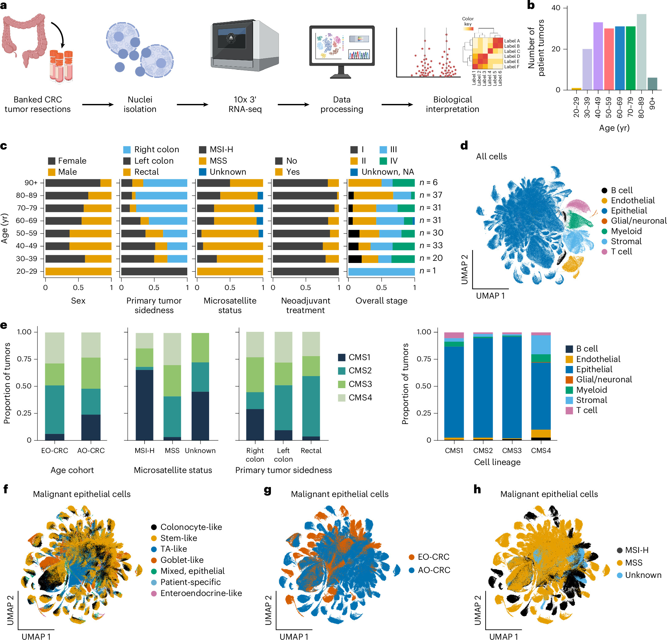
An observational study mapped 189 colorectal tumors across eight decades of life and found age-linked differences in cell proportions within one tumor subtype.
Independent replication
Limit: Tumor location tracks patient age, complicating separation of age effects from location effects
Edited by Boris Djordjevic · AgingResearch.News
An observational study analyzed 189 primary colorectal tumors from patients spanning eight decades of life. Of these, 54 were diagnosed before age 50 and 135 at age 50 or older. Researchers mapped cell types by measuring gene activity in individual cell nuclei, the structures that hold DNA.
In microsatellite-stable tumors, which lack instability in short, repeated DNA sequences, younger age was linked to a higher share of supporting-tissue cells and a lower share of epithelial cells, the type that lines the bowel. The analyses accounted for tumor location, stage, patient sex and treatment before surgery. Researchers confirmed the cell-mix pattern using tissue imaging and machine learning in a separate group of 712 microsatellite-stable tumors: 18 diagnosed before age 50 and 694 at age 50 or older.
Why it matters
Colorectal cancer diagnosed before age 50 is increasing worldwide. Mapping age-associated tumor features may help researchers understand how cancers arising earlier in life differ biologically from those diagnosed later.
Caveats
The observational design cannot show that age caused the cell-mix differences. These findings concerned microsatellite-stable tumors, and the separate validation group included only 18 tumors diagnosed before age 50.
From the paper
- Result
… with more left-sided tumors (Fisher’s exact test, two-sided; odds ratio (OR) 3.61, 95% confidence interval (CI) 1.85–7.04, P = 0.0002) and stage IV tumors (OR 2.55, 95% CI 1.23–5.27, P = 0.017) in EO-CRC compared to AO-CRC.
- Limitation
First, many of the reported findings should be considered hypothesis-generating and warrant validation in future studies.
Quoted word for word from the full text on nature.com.
- Resources: datasets, atlases and tools
- Single-cell omics
- Average-onset colorectal cancer
- Colorectal cancer
- Early-onset colorectal cancer
- Humans
The paper
Broad Institute; Harvard University
Nature Medicine, Oct 29, 2026, Open access


