Lower bone density on lung scans linked to spine fractures
Over two years, spine fractures occurred in 0.9% of participants with normal bone mass, 2.0% with low bone mass and 5.9% with osteoporosis.
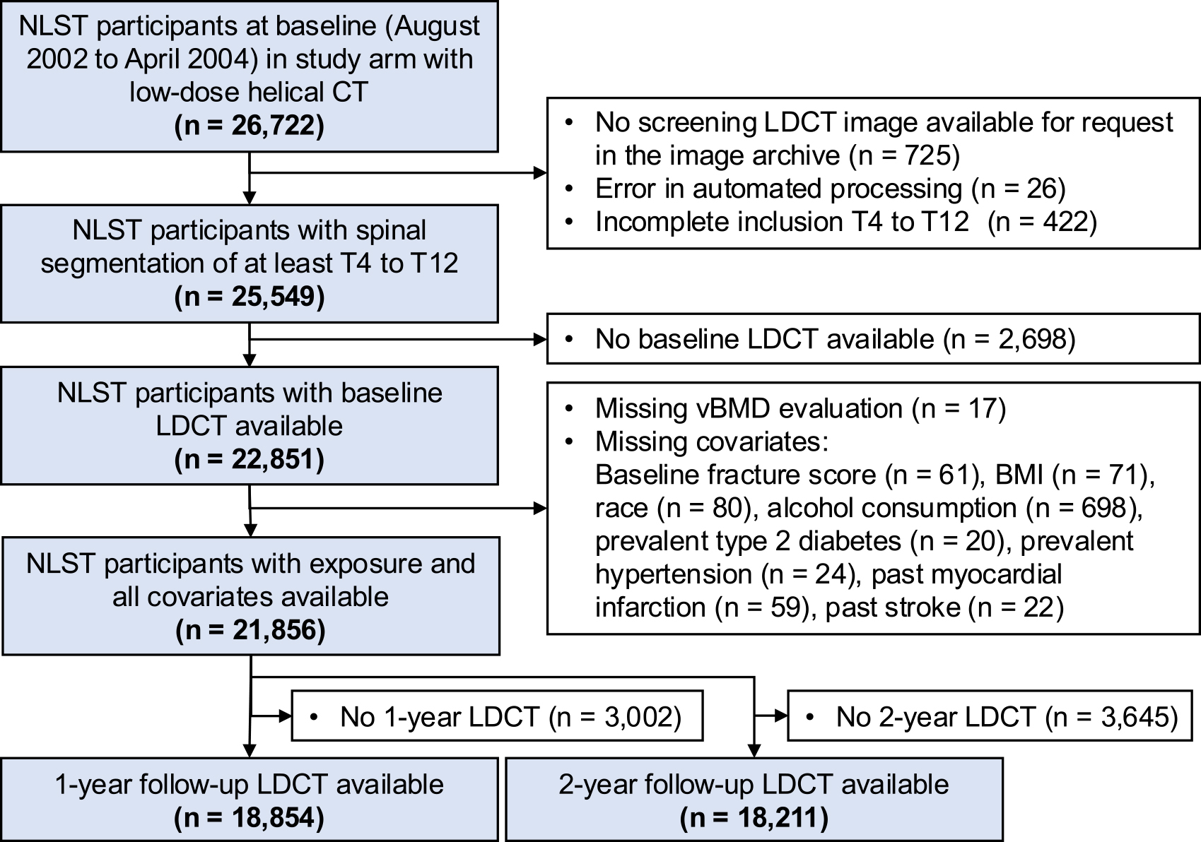
21,856 participants, multicenter, registered NCT00047385
Limit: The fracture analysis excluded participants without two-year scans
Edited by Boris Djordjevic · AgingResearch.News
An observational analysis included 21,856 people aged 55–74 who smoked heavily, including 12,854 men. Researchers analyzed low-dose computed tomography (CT) scans from the National Lung Screening Trial. Software automatically measured spinal bone mineral density—the amount of mineral in a given volume of bone—and identified spine fractures.
Among 18,211 participants with two-year scans, low bone mass and osteoporosis, a condition of weakened bones, were associated with more new spine fractures than normal bone mass. The association remained after adjustment for demographic factors, available health conditions and existing spine fractures. Among 21,447 participants with completed follow-up, the median follow-up was 6.65 years. Bone density in the osteoporosis range was associated with a higher rate of death in men, with an adjusted hazard ratio of 1.24.
Why it matters
The study addresses whether lung cancer screening can also inform bone health care in older adults. The authors noted that two-year fracture incidence in participants with osteoporosis exceeded the 5% treatment threshold in current osteoporosis guidelines.
Caveats
This secondary analysis used only the CT arm of a randomized lung-screening trial and cannot show that low bone density caused fractures or death. It was limited to people aged 55–74 who smoked heavily, and the two-year fracture analysis included only participants with two-year scans.


