Tiny vesicles spread senescence in lab-grown human cells
In lab-grown human fibroblasts, tiny vesicles from senescent cells prompted dividing cells to halt division and activate a DNA damage response.
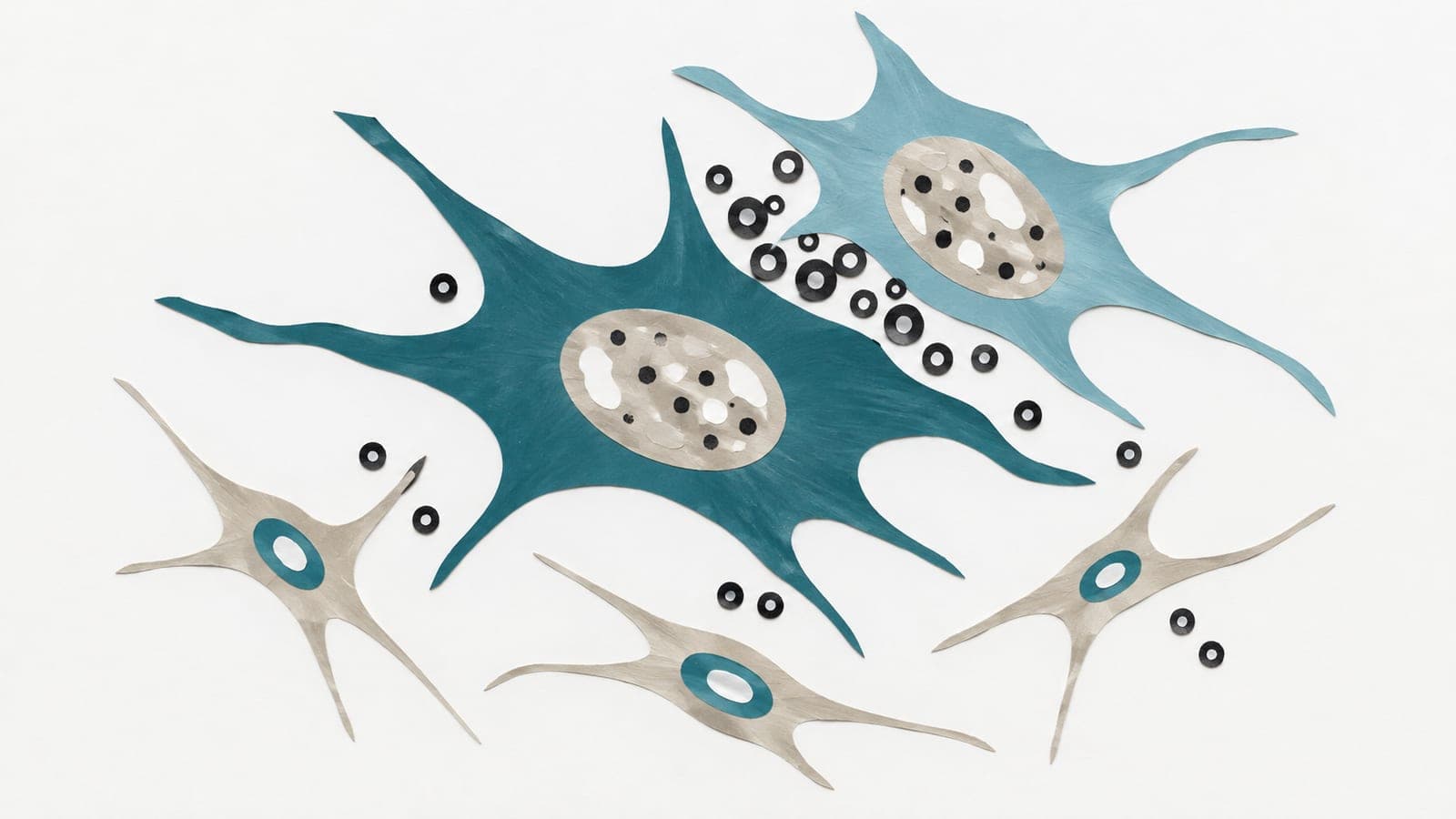
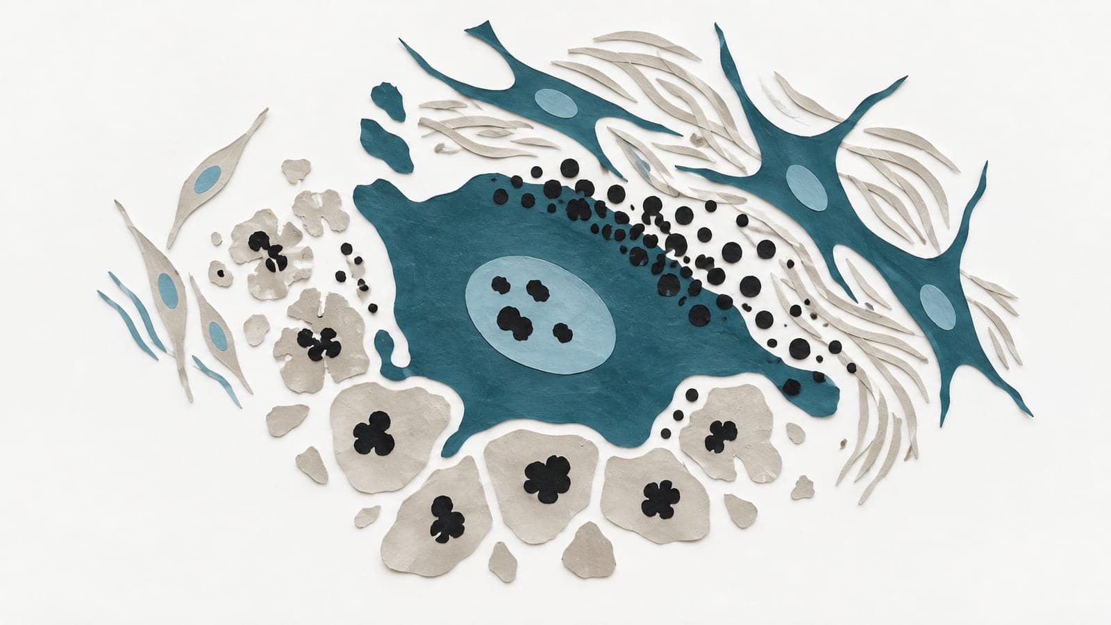
In experiments using human primary fibroblasts grown in the lab, researchers investigated how senescent cells, which permanently stop dividing, communicate with nearby cells. Fibroblasts engineered to produce the cell-cycle inhibitor p16INK4A released significantly more small extracellular vesicles—tiny fluid-filled packages—than proliferating cells. When researchers collected these vesicles and applied them to proliferating fibroblasts, the recipient cells halted division, produced more p16INK4A, and activated a DNA damage response. This transmission occurred without inducing the classical inflammatory signals interleukin-6 and interleukin-8. Similar vesicle-driven transmission occurred in two cellular models of aging: cells expressing progerin and cells with telomere dysfunction. Vesicles isolated from fibroblasts derived from aged donors also induced senescence in fibroblasts from young donors.
Why it matters
Senescent cells accumulate during aging and influence neighbouring tissue environments. Identifying small extracellular vesicles as carriers of these signals may help clarify how cellular aging spreads without relying on a classical inflammatory response.
Caveats
The findings come entirely from cells cultured in the lab, which may not capture how extracellular vesicles behave in living human tissues. The study also relied on specific cellular models of aging that may not represent every way senescence occurs.