MRI morphometry of the popliteus tendon and lateral meniscus in an Iraqi adult cohort
Abstract
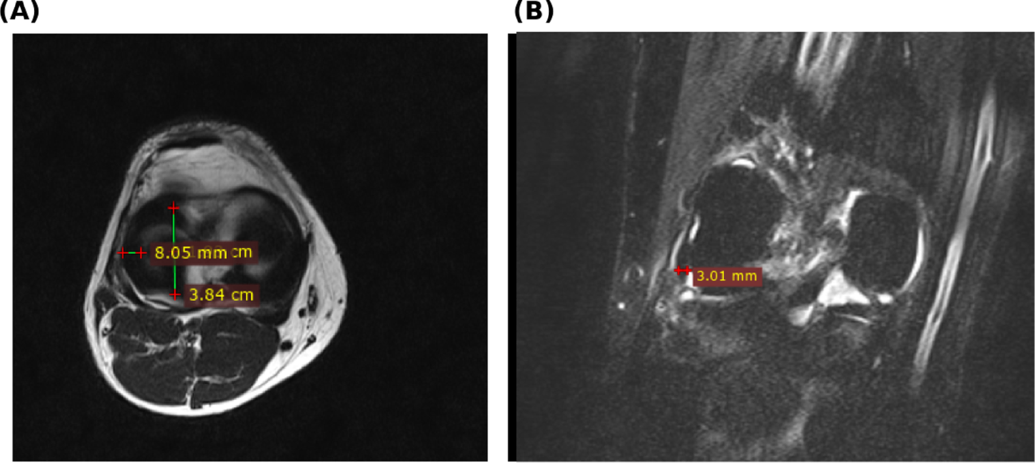
PURPOSE: This study provides sex-stratified and age-related MRI normative values for the popliteus tendon thickness and lateral meniscus width in an Iraqi adult cohort. METHODS: A retrospective study performed at the Medical City Complex, a tertiary-care center in Baghdad. A total of 120 normal adult knees (61 males, 59 females; age 18-66 years) were imaged on a 1.5-Tesla scanner. The coronal fat-suppressed image was used to measure the popliteus tendon, and the axial T2-weighted image was used to measure the width of the lateral meniscus body. The Mann-Whitney U test with false discovery rate correction was used to evaluate sex differences. Associations with age were evaluated using Spearman's rank correlation. RESULTS: Popliteus tendon thickness was 3.61 ± 0.33 mm in males and 2.77 ± 0.23 mm in females (p < 0.001; r = 0.84). Lateral meniscus body width was 8.35 ± 0.47 mm and 7.48 ± 0.48 mm, respectively (p < 0.001; r = 0.69). Sex remained independently associated with both measurements after adjustment for age (Popliteus tendon 0.762 mm, 95% CI 0.677-0.847; lateral meniscus body width 0.809 mm, 0.641-0.977; both p < 0.001). Both structures declined with age (ρ = - 0.491 and - 0.353), with a steeper decline observed for the popliteus tendon (p = 0.037). CONCLUSION: Preliminary sex-stratified normative values for the popliteus tendon and lateral meniscus, obtained through MRI, are reported for an Iraqi adult cohort. Both anatomical structures exhibit significant sexual dimorphism and age-related decline, underscoring the importance of accounting for sex and age when interpreting their morphometry.
The paper
University of Baghdad
Surgical and Radiologic Anatomy, 12 Aug 2026, Open access, accepted manuscript