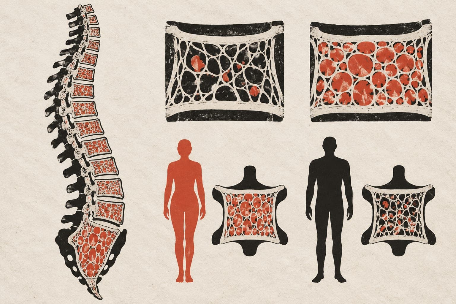
Metabolism and bone marrow : Imaging of physiological and pathological processes
Abstract
CLINICAL/METHODICAL ISSUE: Human bone marrow and its metabolism-particularly the composition of bone marrow adipose tissue (BMAT)-are subject to physiological changes throughout the lifespan. However, they are also involved in and altered by pathological processes. Modern techniques of quantitative magnetic resonance imaging (MRI) now enable an objective and quantitative evaluation of such processes. STANDARD RADIOLOGICAL METHODS: Standard radiological procedures consist of T1- and T2-weighted MRI of the bone (marrow). METHODICAL INNOVATIONS: Methodological innovations consist of chemical shift encoding-based (CSE) water-fat imaging and proton (¹H) MR spectroscopy (MRS). PERFORMANCE: In particular, quantification using the proton density fat fraction (PDFF) is of relevance for both physiological and pathological changes of BMAT. ACHIEVEMENTS: The determination of the PDFF-particularly by means of (¹H)-MRS-is increasingly being used for the evaluation of BMAT, but it remains a non-standard procedure. PRACTICAL RECOMMENDATIONS: Knowledge of the typical signal characteristics of BMAT is essential for reliably discriminating physiological from pathological changes-both in terms of visual image reading and quantification.
The paper
University of Freiburg; TUM Klinikum
Radiologie, 15 Jul 2026