Computer model flags very fragile bones on adults’ X-rays
Against bone density scans, internal testing yielded specificity of 0.93 for correctly ruling out very high fragility and sensitivity of 0.53 for detecting it.
Our read
Biomarkers- Studied:
- Cohort study of 11,138 people (Independent replication, multicenter)
- Stake:
- Five authors work for Medimaps Group and one holds shares in it.
- In the record:
- One of 8 papers on deep learning model on record: 6 with results in people.
Editor of record: Boris Djordjevic · AgingResearch.News
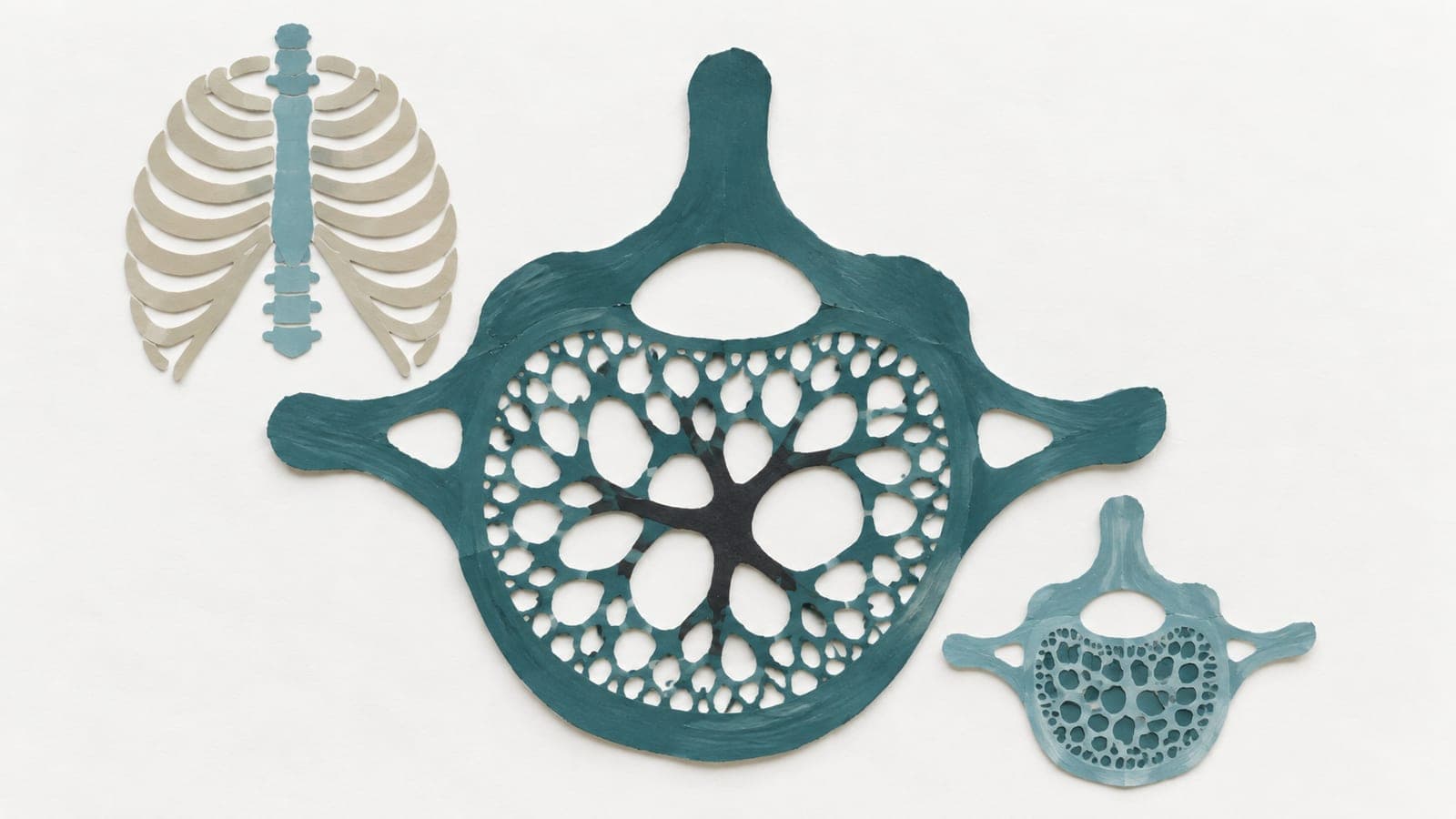
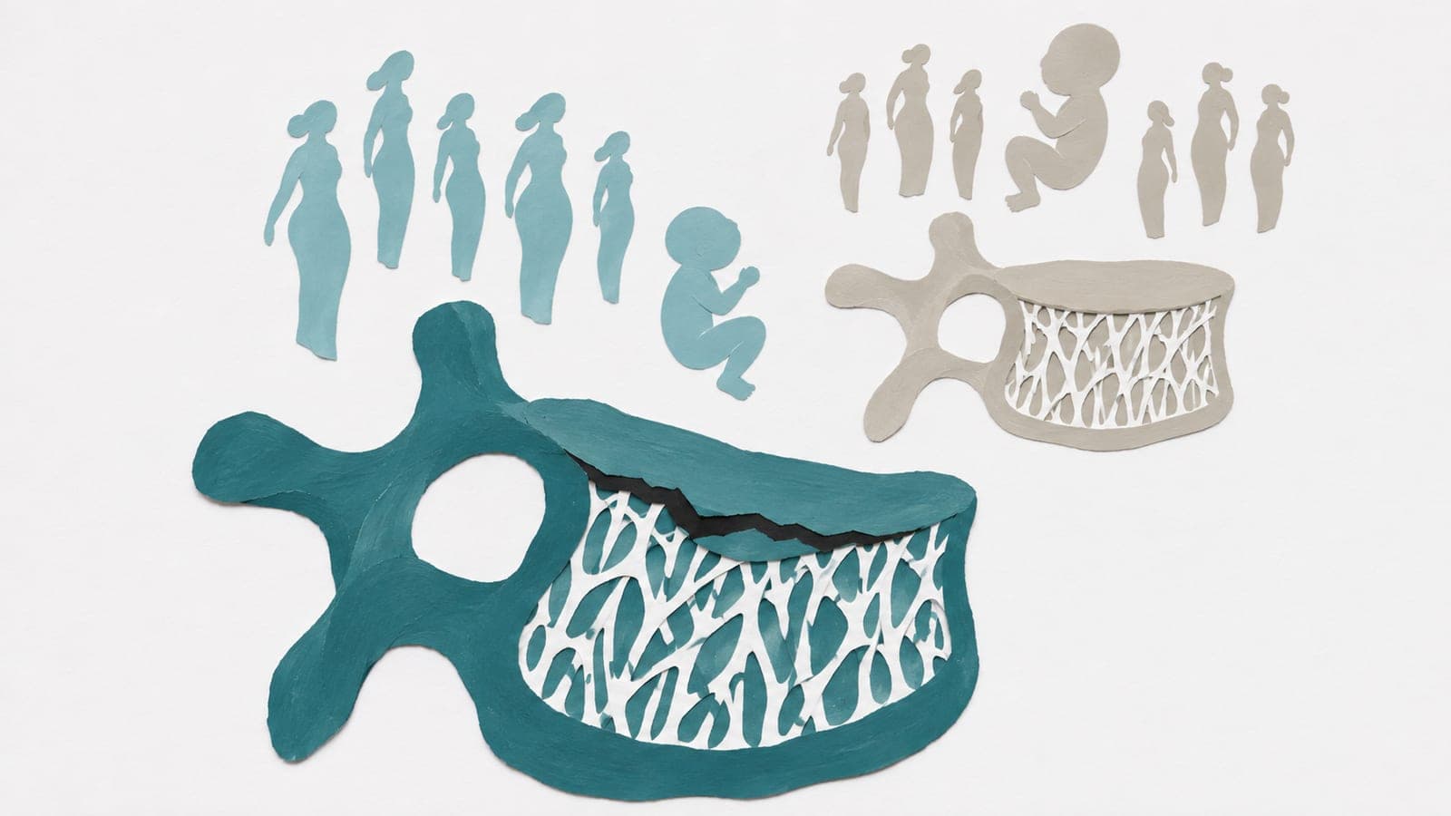
Routine X-rays and bone density scans from 11,138 adults aged 20 or older at five European and US sites were analyzed in a retrospective cohort study. Average ages across sites ranged from 62.5 to 69.6 years. The artificial intelligence model looked for very high bone fragility, defined as osteoporosis—abnormally low bone density—combined with a degraded score reflecting internal bone structure. Each X-ray showed the lower spine and had a corresponding bone density scan within six months.
The team trained the model on Italian and Austrian data and tested it on separate data from those countries, Slovakia and the US. Internal testing yielded specificity of 0.93, measuring correct exclusion of very high fragility, and sensitivity of 0.53, measuring detection. External specificity was 0.88 in the European group, 0.94 in White Americans and 0.96 in non-White Americans.
Why it matters
Osteoporosis screening aims to identify people at risk of fragility fractures. The authors propose using images already taken in routine care as another opportunity to screen.
Caveats
The model identified a scan-defined fragility category, not future fractures. All participants already had bone density scans, so performance in people without prior testing remains untested.
The paper
Centre Hospitalier Universitaire Vaudois
EClinicalMedicine, May 13, 2026, Open access
Comments
Be the first to comment.