Brain age profiles capture disease-linked patterns in people
Adding imaging profiles improved scores for distinguishing disease groups by 0.018–0.060 compared with a baseline using age, brain age deviation, sex and imaging center.
Limit: Recorded diagnoses can miss subclinical disease and misdate biological onset
Edited by Boris Djordjevic · AgingResearch.News
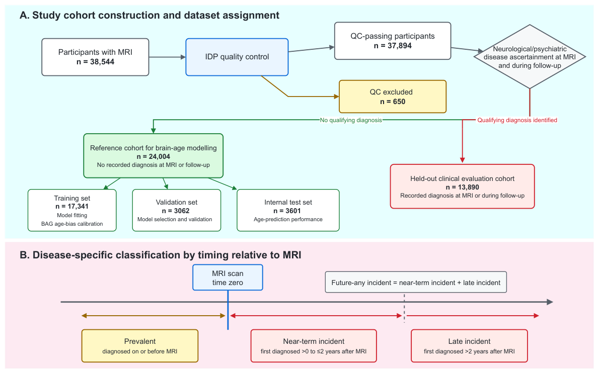
In an observational analysis of 37,894 UK Biobank participants, researchers developed a model that predicted age from several types of brain imaging measurements. They evaluated it using separate reference and clinical groups. The model also produced individual profiles describing the imaging patterns behind each age prediction.
The brain age gap is the difference between predicted and actual age. Participants with nearly identical gaps on a standardized scale had different imaging profiles. Adding these profiles to a baseline model containing age, standardized brain age gap, sex and imaging center improved its ability to distinguish groups in four disease comparisons. The gains persisted after researchers matched participants by age and standardized brain age gap.
Why it matters
For research on brain aging, the authors propose these profiles as a way to examine disease-related imaging differences beyond a single age estimate.
Caveats
This observational model evaluation did not establish clinical diagnostic usefulness. The abstract does not identify the four disease comparisons or report participants’ ages.
From the paper
- Participants
The reference cohort comprised 24,004 participants and was divided into training, validation, and test sets.
Results, 3.1. Study Population and Brain Age Model Performance
- Limitation
The evaluation was internal to UK Biobank participants aged 44–83 years, without independent validation in other populations.
Quoted word for word from the full text on mdpi.com.
The paper
Northwestern Polytechnical University
Biology, Oct 22, 2026, Open access, CC BY


