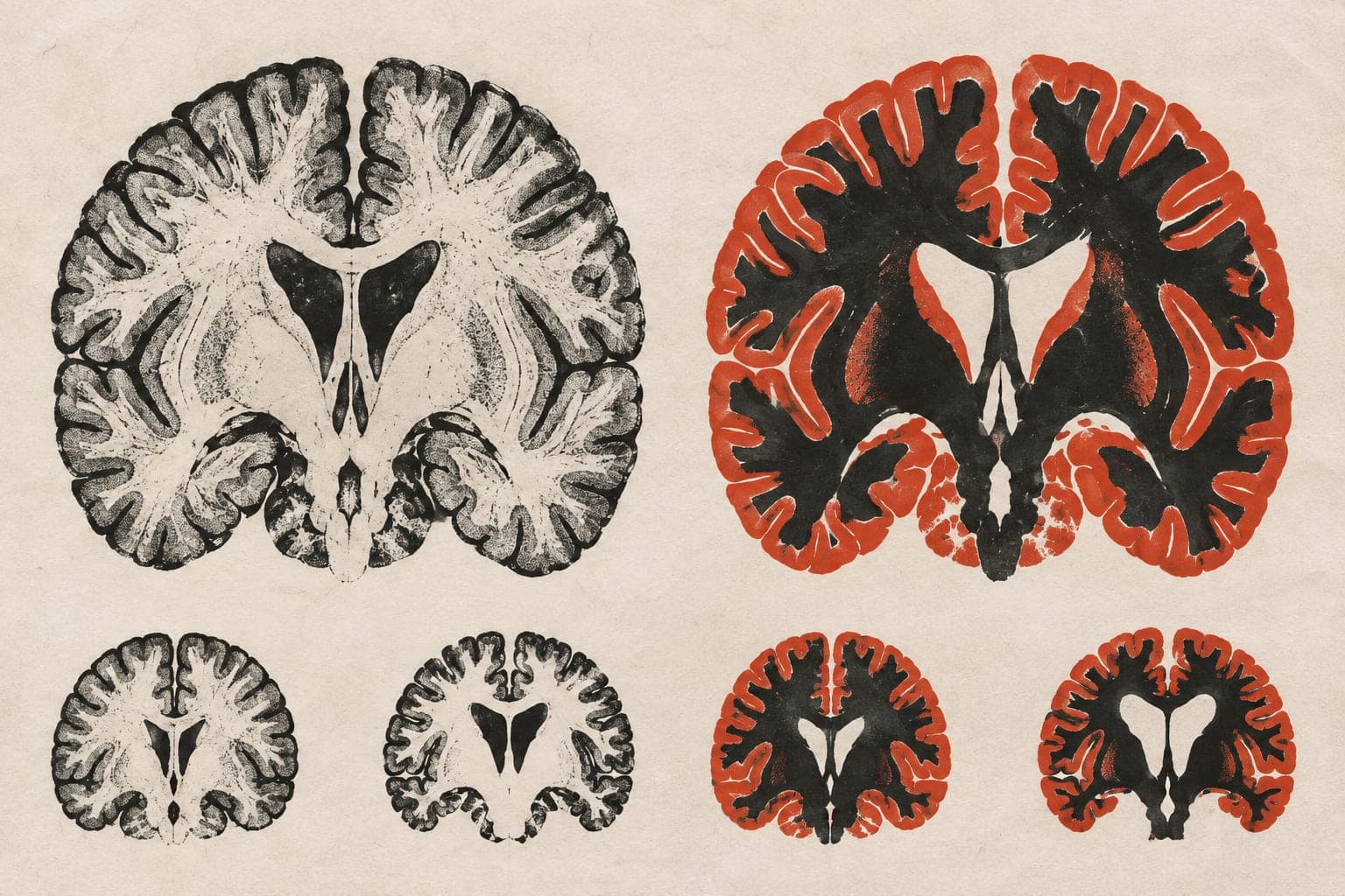
Model estimates brain aging speed from local MRI changes
Tested on large-scale scan datasets, the computational framework combined regional brain shape changes to track aging speed and spot abnormal patterns in disease.
Medical Image Computing and Computer-assisted Intervention
Researchers tested a computational framework on large-scale datasets of brain scans taken over time, including scans from diseased populations. Magnetic resonance imaging (MRI) can show brain structure, but many tools assess brain age at only a single visit rather than measuring the speed of aging over time. Existing longitudinal tools also rely heavily on image textures and can overlook how different brain areas age at different rates. To address this, the team analysed structural shape changes between pairs of scans. Their method calculates the pace of aging across local brain patches and then combines those regional estimates into a single overall aging speed. The approach predicted aging speed more accurately than existing methods and improved the identification of abnormal aging patterns in diseased groups.
Why it matters
Tracking how quickly different brain regions change over time may help researchers better characterise cognitive decline and evaluate risks for neurodegenerative disease.
Caveats
The findings come from computational modelling of scan data, and the abstract does not report the specific diseases, sample sizes, or demographic details of the datasets analysed.
The paper
University of Pennsylvania
Medical Image Computing and Computer-assisted Intervention · 24 Sep 2026