Retinal scan model identified existing brain blood vessel disease
Detection scores were 0.95 in internal testing and 0.94 in external testing, where higher scores mean better separation of people with and without disease.
Our read
Biomarkers- Studied:
- Cohort study of people assessed for brain blood vessel disease, against people without disease
- What changed:
- The retinal model detected existing brain blood vessel disease with an area-under-the-curve score of 0.94 in external testing
- The catch:
- The main detection scores measure existing disease, not future stroke risk
Editor of record: Boris Djordjevic · AgingResearch.News
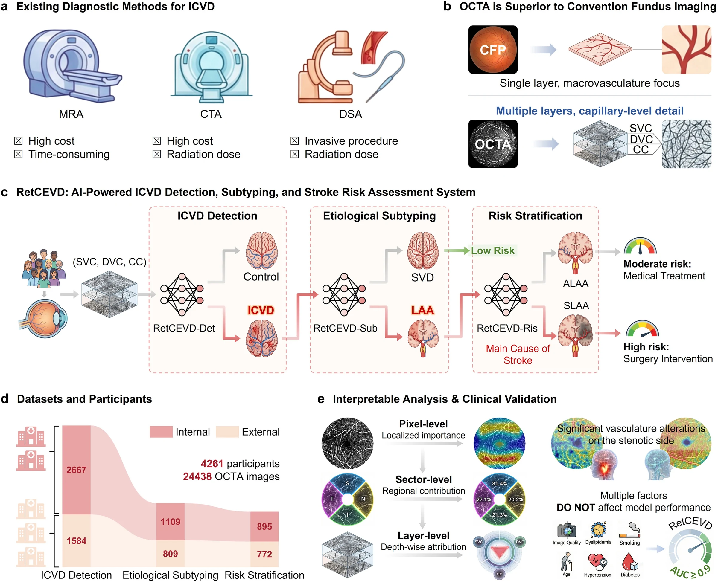
People in a multicenter observational study contributed 24,438 retinal images to train and validate RetCEVD, a computer model for assessing disease in the brain’s blood vessels. Noninvasive scans mapped tiny blood vessels in the retina, the light-sensitive layer at the back of the eye.
The model detected existing disease with area-under-the-curve scores of 0.95 in internal testing and 0.94 in external testing. This measure describes how well a test separates groups. It also distinguished disease involving plaque buildup in large arteries from small-vessel disease, and classified whether people with large-artery disease currently had symptoms. Supplementary two-year follow-up in people with symptom-free large-artery disease yielded a score of 0.71 for subsequent strokes.
Why it matters
Stroke prevention depends on identifying people at risk before neurological symptoms appear. The authors suggest retinal imaging may help identify people who need confirmatory brain-vessel imaging and specialist assessment.
Caveats
The main tests assessed disease already present, so their scores should not be read as predictions of future stroke. The two-year follow-up provided supplementary evidence about later events.
Comments
Be the first to comment.