High blood pressure duration linked to brain tissue differences
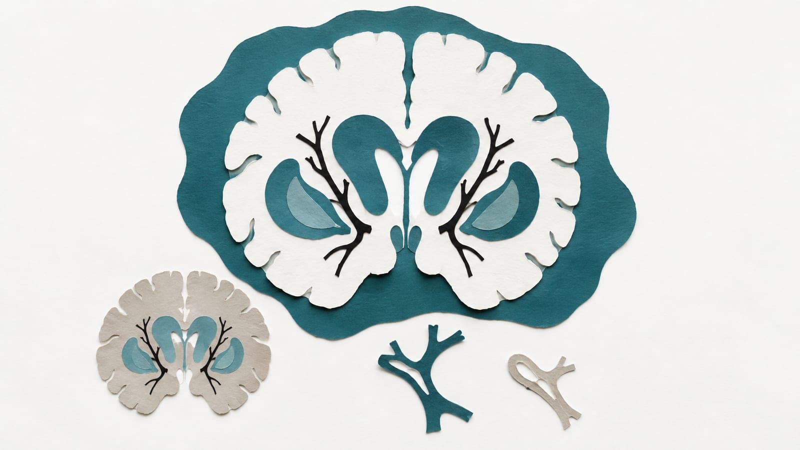
A cross-sectional study of 487 people with high blood pressure and 165 controls found region-specific differences in grey matter volume across disease-duration groups.
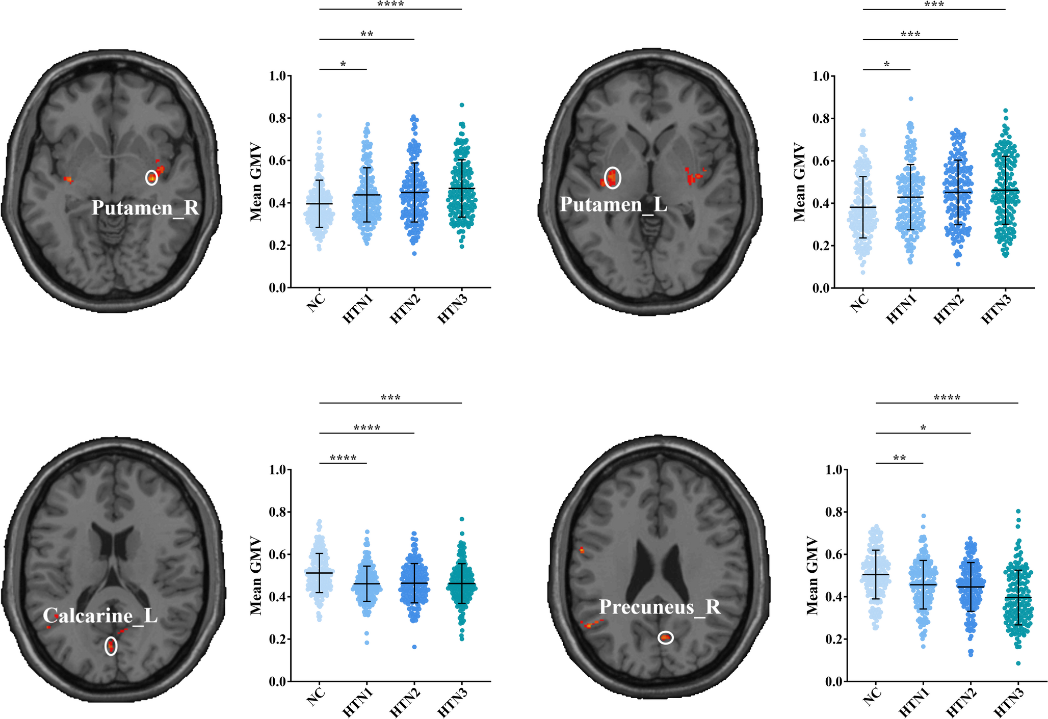
In a cross-sectional observational study, researchers analysed brain scans and cognitive tests from 487 people with high blood pressure and 165 people with normal blood pressure. They grouped those with high blood pressure by duration: four years or less, five to 11 years, and 12 years or more.
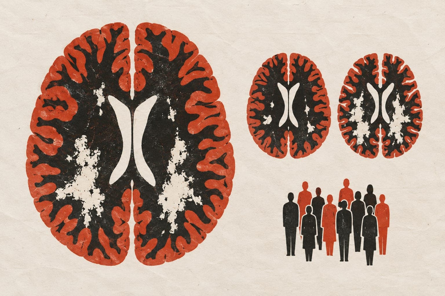
They measured grey matter volume, the amount of brain tissue rich in nerve cell bodies. They adjusted analyses for age, sex, volume inside the skull and, where appropriate, education. Longer disease duration was linked to lower volume in some regions and higher volume in others after correction for multiple comparisons. Only greater volume in one region remained linked to higher cognitive test scores after correction. The regional differences did not fit a uniform pattern of lower volume with longer disease duration.
Why it matters
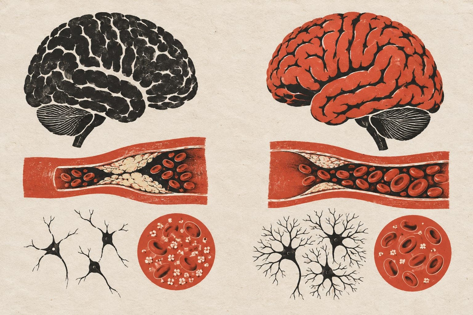
High blood pressure is a modifiable risk factor for cognitive decline. Its duration may be important when evaluating brain structure in studies of cognitive health.
Caveats
This cross-sectional study compared different people rather than tracking changes within individuals. It could not establish cause and effect or show how these brain differences developed over time.
- Hypertension
- Gray matter volume in the bilateral putamen and right insula
- Gray matter volume in the right rectus, left middle temporal, and left angular gyri
- Left thalamic gray matter volume
- MoCA score
- Humans
The paper
Shanghai University; Capital Medical University Affiliated Beijing Friendship Hospital
Brain Imaging and Behavior, 12 Oct 2026