Post-menopausal women show larger brain regions in UK study
Brain scans from 4,098 women revealed greater gray matter volume and cortical thickness in specific regions among post-menopausal compared with pre-menopausal women.
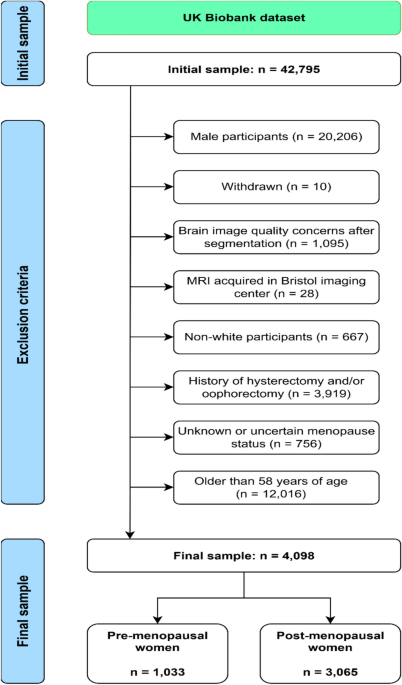
In an observational study, researchers analysed brain scans from 4,098 women, including 1,033 pre-menopausal and 3,065 post-menopausal participants, from the UK Biobank. The team evaluated gray matter volume, cortical thickness, and cortical gyrification, which refers to brain folding. Compared with pre-menopausal women, post-menopausal women showed greater gray matter volume primarily in the right temporal lobe, including the amygdala and hippocampus, extending to the left temporal lobe and occipital areas. Post-menopausal women also exhibited greater cortical thickness in the frontal lobe and the supramarginal gyrus. Cortical folding did not differ between the two groups. The findings remained consistent after accounting for age differences, peri-menopausal status, and other potential confounding factors.
Why it matters
The structural differences overlap considerably with brain regions affected early in Alzheimer's disease, such as the medial temporal lobe. The researchers note that greater volume and thickness may reflect compensatory mechanisms, speculatively aligning with a biphasic model of structural changes in Alzheimer's disease.
Caveats
Because this study analysed cross-sectional brain scans taken at one moment in time, it cannot show how brain structure changes within individual women across menopause. Longitudinal studies tracking the same women over time are needed to uncover the mechanisms behind these differences.
The paper
George Mason University; Instituto de Salud Carlos III
Brain Structure & Function, 9 Oct 2026