Membrane attack complex deposits on pericytes in human eyes
Single-cell analysis of four aged human donor eyes revealed complement membrane attack complexes concentrated primarily on pericytes, smooth muscle cells, and macrophages.
Our read
Mechanisms- Studied:
- Preprint: cross-sectional study of 4 people
- In the record:
- One of 6 papers on CD59 on record: 2 with results in people, 1 in animals.
Editor of record: Boris Djordjevic · AgingResearch.News
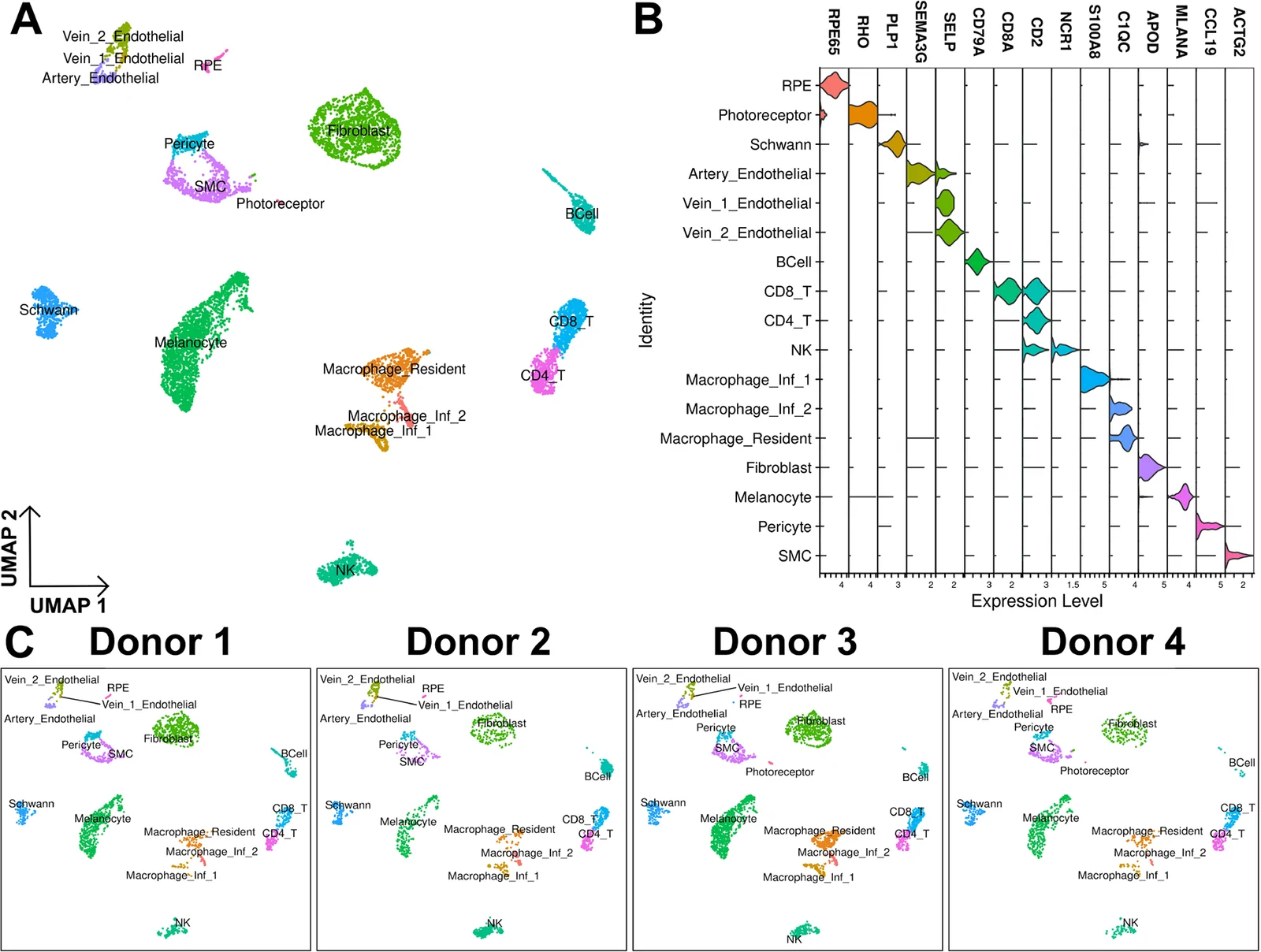
In tissue from four aged human donor eyes, researchers profiled surface complement proteins alongside transcriptomes at single-cell resolution. Using single-cell RNA sequencing combined with barcoded antibodies, the team measured the complement membrane attack complex (MAC) along with complement regulators CD55 and CD59 across retinal pigment epithelium and choroid samples. Surface CD55 and CD59 appeared across various cell types and were more abundant on venous endothelial clusters, correlating with expression of clusterin. MAC was detected on multiple cell types but showed the highest abundance on smooth muscle cells, macrophage-family cells, and pericytes. Immunohistochemistry confirmed MAC deposition on choriocapillaris pericytes.
Why it matters
Identifying cell-specific complement deposition provides insight into how terminal complement activation may drive vascular cell dropout and tissue dysfunction in age-related macular degeneration.
Caveats
The analysis is based on tissue samples from only four human donors, and the study is a preprint that has not yet completed peer review.
The paper
University of Iowa
bioRxiv, Sep 10, 2026, Open access, Preprint, not yet peer-reviewed


Comments
Be the first to comment.