Cell-released particles reduce aging-linked signs in human cells
Stressed human heart-vessel lining cells showed fewer markers of senescence, a state in which cells stop dividing, after 48 hours of particle exposure in laboratory experiments.
Our read
Interventions- Studied:
- Experiments in cells
Editor of record: Boris Djordjevic · AgingResearch.News
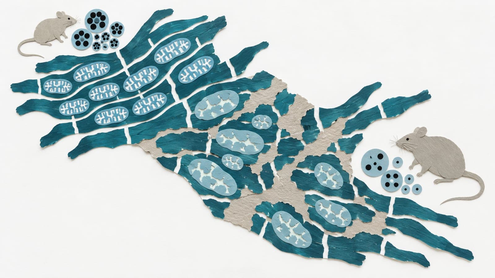
Human cardiac microvascular endothelial cells, which line the heart’s small blood vessels, received extracellular vesicles—tiny cell-released packages—in a laboratory experiment. The vesicles came from support cells grown from human cells reprogrammed into stem cells. After hydrogen peroxide exposure, the endothelial cells received 20 micrograms of vesicle-preparation protein per milliliter for 48 hours.
Vesicles reduced senescence-associated markers and increased the share of living cells and cells showing signs of division. They lowered TXNIP, a protein involved in stress responses. Artificially raising TXNIP increased senescence markers in vesicle-treated cells. In 22-month-old male mice, with five animals per group, seven injections over 21 days increased heart-muscle oxygen saturation and reduced a DNA-damage signal in blood-vessel lining cells compared with saline.
Why it matters
Senescence in blood-vessel lining cells contributes to cardiac aging. The authors concluded that the findings support further investigation of these vesicles in aging of the heart’s small blood vessels.
Caveats
The authors note that changes in these cellular measurements did not establish a lasting reversal of senescence.
From the paper
- Limitation
The transcriptomic dataset included one 10-year-old and two 20-year-old male macaques, restricting age comparisons to descriptive analyses.
Quoted word for word from the full text on mdpi.com.
Comments
Be the first to comment.