Age-linked marks in human brain cells overlap Alzheimer’s changes
An observational analysis of brain tissue from 29 neurologically healthy donors linked age-related changes in non-neuronal cells to molecular patterns associated with Alzheimer’s disease.
Our read
Mechanisms- Studied:
- Cross-sectional study of 29 neurologically healthy human brain donors
- What changed:
- Age-gained brain-cell marks overlapped Alzheimer’s-associated regions at 250 sites, versus fewer than 15 overlaps on average with random fragments
- The catch:
- Chromatin-mark measurements lacked matched input controls to distinguish signal from background
- Why it’s aging research:
- It looks at how biology varies with age or lifespan.
Editor of record: Boris Djordjevic · AgingResearch.News
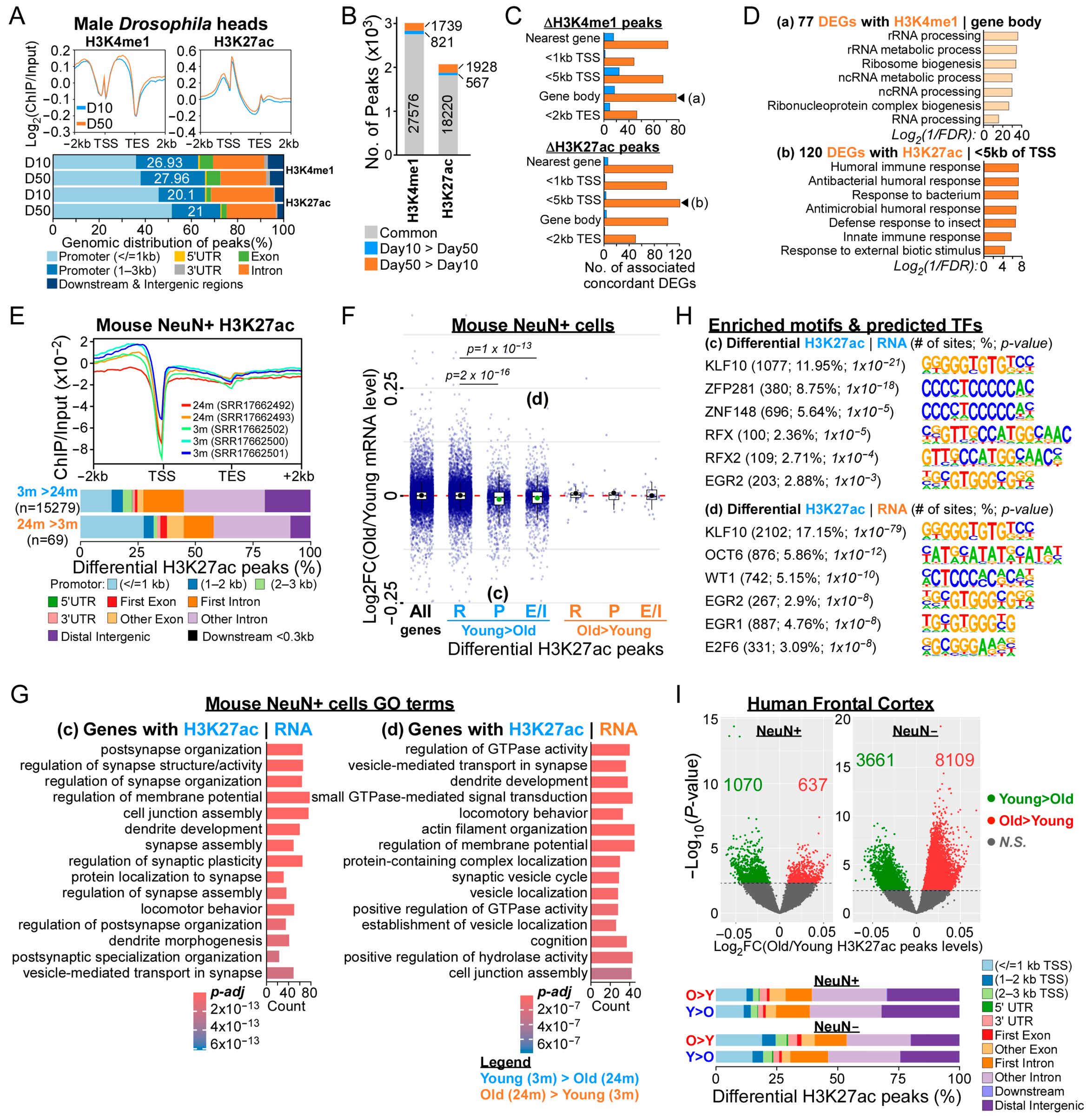
Human frontal cortex cells from 29 neurologically healthy donors were examined after death in an observational analysis. The donors were grouped as 30 or younger, 31–50, and over 50. The researchers compared gene activity with H3K27ac, a chemical tag on DNA-packaging proteins associated with active genes. The wider study also analyzed aging fly heads and mouse forebrain neurons.
Cells other than neurons—the cells that transmit nerve signals—showed more age-associated changes in this tag than neurons did. In these non-neuronal cells, sites where the tag increased with age overlapped Alzheimer’s-associated marks and genetic variants linked to the disease. Genes associated with these age-gained marks also overlapped genes whose activity differed in Alzheimer’s disease.
Why it matters
Gene regulation offers a way to investigate which molecular features of brain aging are shared with Alzheimer’s disease and which are distinct.
Caveats
Postmortem age comparisons could not distinguish marks contributing to disease from changes accompanying normal aging. The mark measurements also lacked matched laboratory controls.


Comments
Be the first to comment.