Protein loss increases bone loss in an animal model
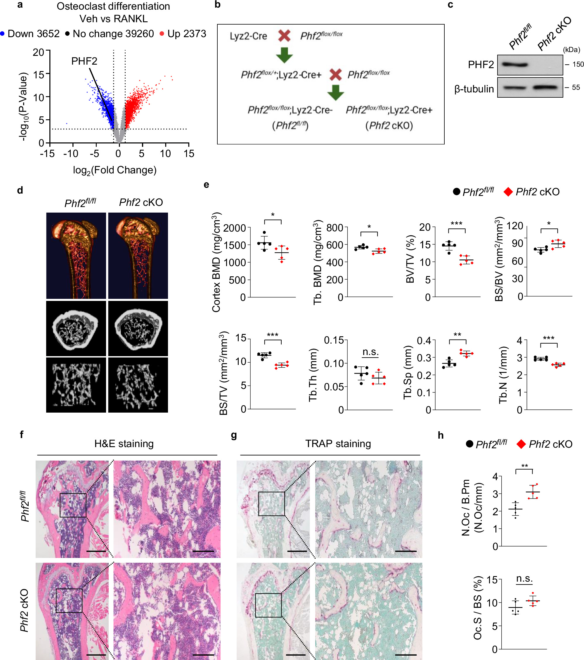
Removing PHF2 from certain bone marrow-derived cells increased bone-resorbing cell formation and deterioration of the spongy tissue inside bones.
Our read
Mechanisms- Studied:
- Experiments in animals with myeloid Phf2 deficiency
- What changed:
- Deleting PHF2 from myeloid cells increased spongy bone loss in animals
- The catch:
- Animal sample sizes are missing from the abstract
- In the record:
- One of 3 papers on Phf2 on record: none yet with results in people.
Editor of record: Boris Djordjevic · AgingResearch.News
In experiments on bone-resorbing cell formation and in animals, researchers identified PHF2 as a protein that restrained osteoclast formation. Osteoclasts are cells that break down bone. Removing the gene for PHF2 from myeloid cells, a family of bone marrow-derived cells, increased osteoclast formation and loss of spongy bone. Enhancing PHF2 function suppressed osteoclast formation.
PHF2 attached to NFATc1, a protein that controls gene activity, and chemically modified it in a laboratory test. The researchers reported that this modification helped mark NFATc1 for breakdown. A change at the modification site stabilized NFATc1 and increased osteoclast formation. In animals whose ovaries had been removed to model postmenopausal osteoporosis, deleting PHF2 from myeloid cells worsened spongy bone deterioration and osteoclast activity.
Why it matters
Postmenopausal osteoporosis involves bone deterioration. The work addresses how controls on bone-resorbing cell formation may matter in this age-related disease.
Caveats
These were laboratory and animal experiments, not a study in people. The abstract does not identify the animal species or report sample sizes.
Comments
Be the first to comment.