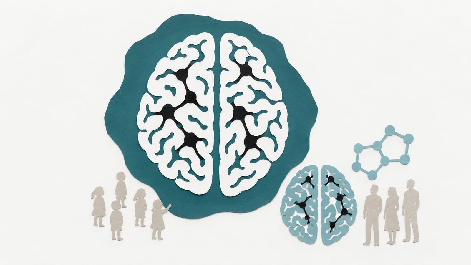
Brain network patterns linked to biology across the lifespan
An observational analysis of resting brain scans from 5,349 people aged 6–80 linked age-related network patterns to cellular and molecular maps.

Cohort study
Limit: Cellular and molecular features were from maps, not measured in participants
Edited by Boris Djordjevic · AgingResearch.News
In an observational study of 5,349 people aged 6–80, researchers compared coordinated brain activity across regions with maps of 49 cellular and molecular features. They used brain scans taken at rest. The maps covered cell types, nerve-cell chemical messengers and features of mitochondria, the energy-producing structures inside cells.
The spatial match between age-related brain network changes and these biological features varied by life stage. Matches with cell types peaked in adolescence, those with chemical messenger systems in mid-young adulthood, and those with mitochondrial features in early adulthood. Modeling of schizophrenia, bipolar disorder, major depressive disorder, autism and attention-deficit/hyperactivity disorder also identified disorder-specific and shared brain network patterns associated with the biological maps.
Why it matters
The framework offers a way to generate hypotheses about which cellular and molecular features may be relevant to brain aging and psychiatric dysfunction.
Caveats
These observational spatial associations do not show that cellular or molecular features caused the brain network differences. The work is a preprint and has not been peer-reviewed.
- Modeling and theory
- Synaptic transmission
- Schizophrenia
- Bipolar disorder
- Major depressive disorder
- Humans
The paper
Tri-Institutional Center for Translational Research in Neuroimaging and Data Science (TReNDS), Georgia State University, Georgia Institute of Technology, and Em
bioRxiv, Oct 26, 2026, Open access, CC BY-NC-ND, Preprint, not peer-reviewed