Hip analysis assumption linked to errors in donated human bones
In younger donors, estimates of dense outer bone area had errors up to 36% compared with high-resolution scans when analysis assumed a fixed bone share.
Limit: Cortical fractions measured bone volume, not mineral mass, understating the fraction required by HSA
Edited by Boris Djordjevic · AgingResearch.News
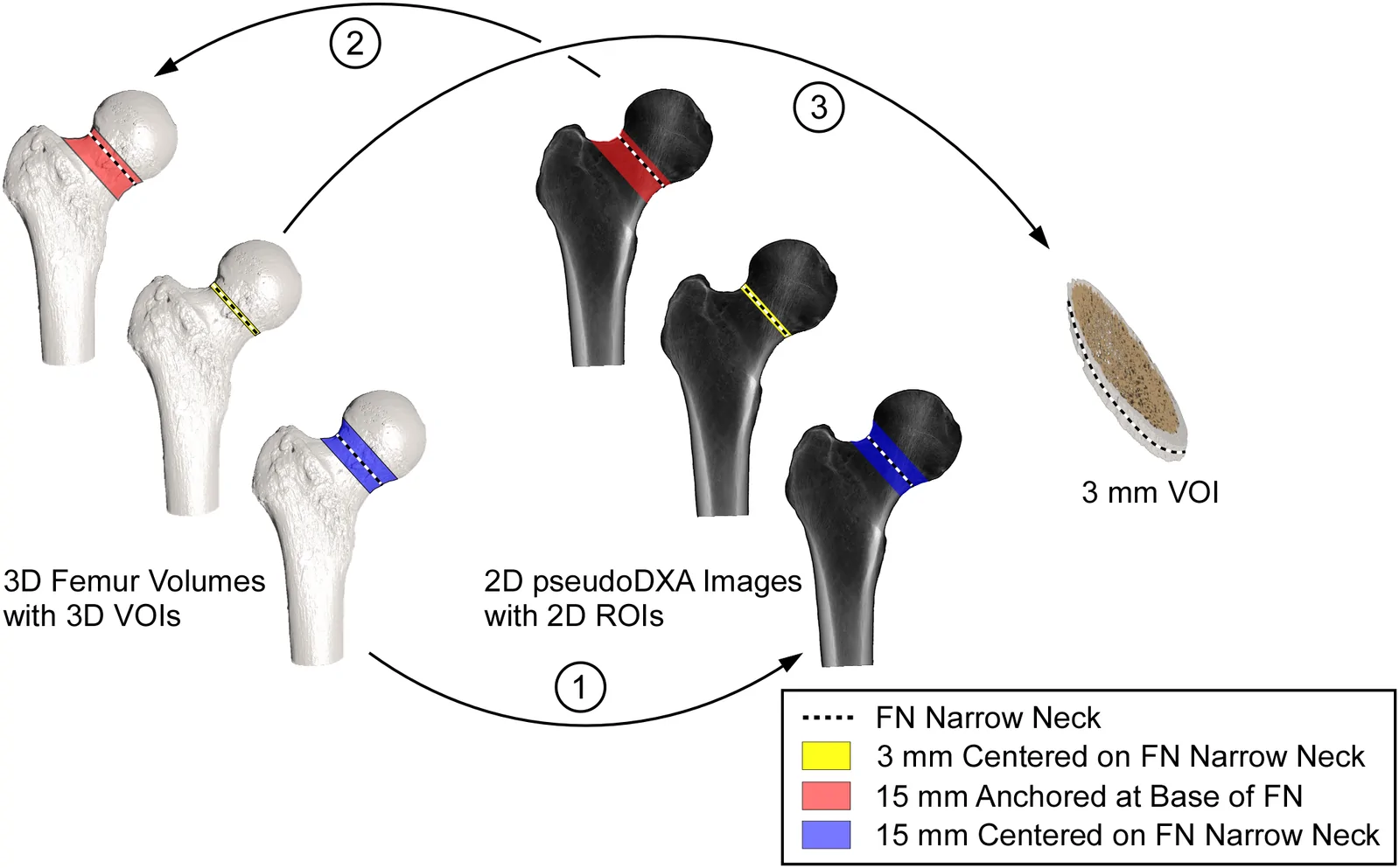
An observational study examined donated thighbones from 32 White female donors aged 24 to over 89 and 30 White male donors aged 18 to 89. Researchers generated simulated bone-density scans from high-resolution three-dimensional images. They tested Hip Structure Analysis, a method for estimating the shape of the femoral neck, the narrow region below the ball of the hip joint.
The method assumed that cortical bone, the dense outer layer, accounted for 60% of bone mineral content. Measured shares varied widely and averaged below that assumption in both sexes. Errors in structural estimates varied by sex and age. Using each bone’s measured share reduced errors in estimates of the outer bone’s cross-sectional area in both sexes.
Why it matters
Hip structure helps inform fracture-risk assessment. The study addresses whether age- and sex-related differences in bone composition need consideration when estimating that risk.
Caveats
The study tested simulated scans of donated bones, not fracture prediction in living people. It included only White donors and excluded samples with known or suspected musculoskeletal disease.
The paper
University of Michigan; University of Colorado Colorado Springs
Frontiers in Endocrinology, Oct 8, 2026, Open access


