Fat-derived protein worsened signs of vessel aging in mice
In experiments with 24 young male mice over 12 weeks, an antibody against asprosin reduced signs of chemically induced blood vessel aging.
Our read
Mechanisms- Studied:
- Experiments in animals
Editor of record: Boris Djordjevic · AgingResearch.News
Twenty-four eight-week-old male C57BL/6 mice were randomly assigned to four groups in a 12-week experiment. The researchers tested asprosin, a protein released by fat tissue, and an antibody against it. Some mice received D-galactose, a chemical used to induce signs of accelerated aging.
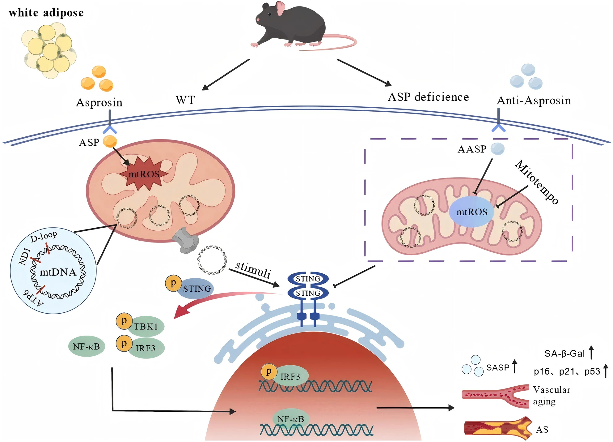
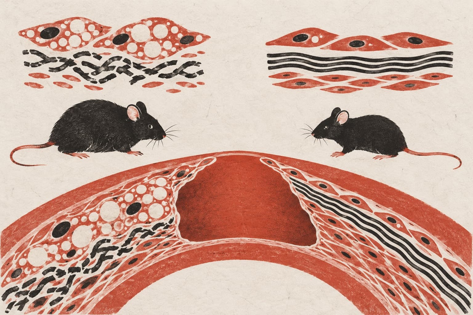
Asprosin increased senescence—a state in which cells stop dividing—in the cells lining blood vessels and worsened their function. The antibody reduced these changes in mice given D-galactose. Mice genetically engineered to lack asprosin in fat tissue also had fewer signs of blood vessel aging. In human umbilical vein lining cells grown in the lab, asprosin increased damaging reactive molecules in mitochondria, the cells’ energy-producing structures. It also caused mitochondrial DNA leakage and activated an immune signaling pathway. Suppressing this pathway or those reactive molecules eased cell senescence.
Why it matters
Aging of the cells lining blood vessels contributes to age-related vascular diseases. The authors suggest that asprosin could be a target for interventions against these diseases.
Caveats
The authors note that most mechanistic experiments used chemically induced aging models and lab-grown cells, leaving open whether the pathway works similarly during natural aging.
From the paper
- Limitation
First, although we observed elevated circulating ASP levels in aged mice, the mechanistic experiments were primarily performed using D-gal-induced senescence models and cultured endothelial cells.
Quoted word for word from the full text on onlinelibrary.wiley.com.
The paper
School of Pharmacy Jiangxi Medical College
Aging Cell, Nov 15, 2026, Open access, CC BY
Comments
Be the first to comment.