Cell-to-cell signals help build artery plaque caps in mice
In mice carrying a blood-cell mutation, activating an immune-cell protein promoted plaque cap formation through signals between immune cells and connective-tissue cells.
Our read
Mechanisms- Studied:
- Preprint: experiments in mice with low-level Jak2V617F clonal hematopoiesis
- What changed:
- Deleting the signal receptor in fibroblast-enriched support cells abolished the plaque cap increase from TREM2 activation
- The catch:
- Plaque structure was measured rather than health benefit
Editor of record: Boris Djordjevic · AgingResearch.News
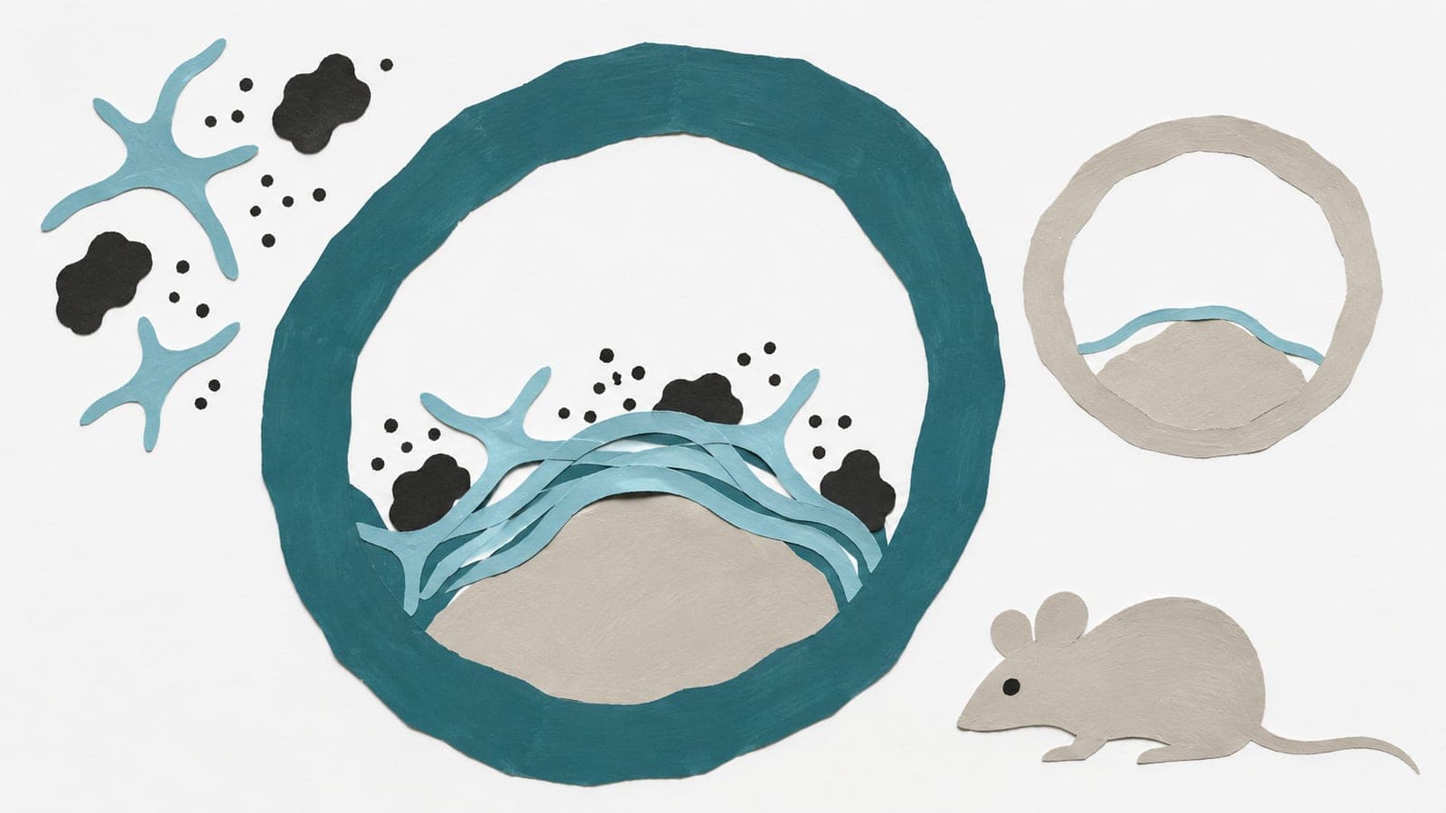
Mice carrying the Jak2V617F mutation at low levels modeled artery plaque disease and clonal hematopoiesis, the expansion of blood cells with a shared mutation. In an animal experiment, they received 4D9, which activates TREM2, a protein on immune cells.
TREM2 activation increased production of PDGFB, a signaling protein, in macrophages, a type of immune cell. It also promoted formation of fibrous caps, the tissue covering plaques. Deleting the PDGFB gene in targeted immune cells markedly weakened this cap response. Deleting the gene for its signal-receiving protein in a group of support cells rich in fibroblasts—cells that make connective tissue—abolished the cap response. That deletion did not significantly change total plaque area or the area of its dead center.
Why it matters
Age-related artery disease involves inflammation and tissue remodeling. The work addresses how communication between immune cells and connective-tissue cells may coordinate these processes within plaques.
Caveats
The experiments measured plaque structure in genetically altered mice, leaving open whether this pathway would offer a health benefit in people. The report is a preprint, not yet peer-reviewed.
The paper
Columbia University Irving Medical Center
bioRxiv, Nov 11, 2026, Open access, CC BY-NC-ND, Preprint, not yet peer-reviewed

Comments
Be the first to comment.